
COVID AND CANCER DETECTION – MEDICAL REPORT
Lymph Node Findings on PET: Cancer or COVID Vaccine?
— Post-jab radiotracer uptake “could prompt unnecessary biopsies and treatments”
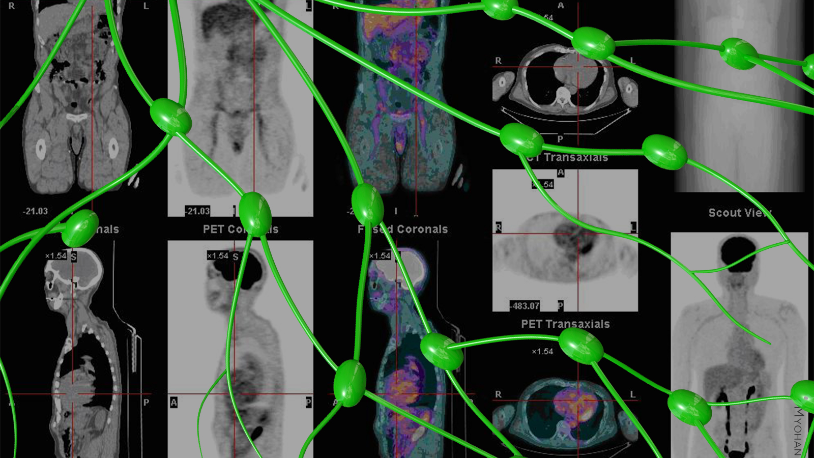
Positron emission tomography (PET) imaging of cancer patients after they received COVID-19 vaccinations showed abnormal radiotracer uptake in lymph nodes, researchers reported.
The retrospective study demonstrated that about 10% of patients, who had no visible axillary nodal uptake on PET imaging performed pre-vaccination, exhibited positive axillary lymph nodes after COVID-19 vaccination, according to Jason Young, MD, of the Mayo Clinic in Rochester, Minnesota, and colleagues.
“The decision to proceed with PET imaging after recent COVID-19 vaccination should take into account the evolving knowledge of the pitfalls that such vaccination can cause on PET, as well as patient-specific factors such as the cancer type, cancer stage, and the urgency of the clinical decisions to be made based on the PET finding,” the authors wrote in the American Journal of Roentgenology.
Observed alterations “could prompt unnecessary biopsies and treatments unless appropriately recognized by interpreting physicians,” they stated.
FDG-avid reactive lymphadenopathy has been reported in patients following their shots with the Pfizer and Moderna COVID-19 vaccines. Young and colleagues noted that systematic analyses of COVID-19 vaccination-induced changes on PET have been limited, particularly the impact of COVID-19 vaccination performed with radiotracers other than FDG, such as choline C-11.
Their study included 67 patients (mean age 76) who underwent PET exams from Dec. 14, 2020 to March 10, 2021, after receiving either the Pfizer or Moderna COVID-19 vaccine. These patients had also undergone pre-vaccination PET without visible axillary node uptake.
PET was performed a median of 13 days after vaccination in 44 patients who had received one vaccine dose, and 10 days in 23 patients who had received two doses.
Positive axillary lymph node uptake was seen in seven (10.4%) patients, four of whom had FDG-PET exams (out of 54 patients), and three of whom had choline C-11 exams (out of 13). Ipsilateral deltoid uptake was observed in 14.5% of patients with documented injection laterality, including three of the seven patients with positive axillary lymph nodes.
All of the examinations showing positive axillary lymph nodes were performed within 24 days of vaccination.
In a separate earlier American Journal of Roentgenology study, Lacey J. McIntosh, DO, MPH, of the University of Massachusetts Medical School in Worcester, and colleagues suggested that, based on their institution’s experience with COVID-19 vaccination-related uptake on FDG PET/CT, the exam should be performed at least 2 weeks after vaccination — ideally 4-6 weeks if the exam is not urgent — in patients with a cancer for which interpretation could be impacted by the vaccine.
“At baseline these PET scans are a comprehensive way of telling where a cancer is, and how to target and treat it,” McIntosh told MedPage Today. “The entire treatment plan is often based on results of this scan.”
McIntosh said that in some cases it is relatively simple to identify COVID-19 vaccination-related uptake based on vaccination history of the patient, and the natural history of the cancer clinicians are trying to evaluate. In other cases, management could depend on whether the finding is clinically relevant or irrelevant. “If the patient has a widespread cancer and has axillary lymph node involvement, it’s not going to matter whether they are involved due to the cancer or because of the vaccination,” she pointed out. “It’s not really going to change the patient’s staging or course of treatment.”
“Situations where we can really run into trouble are the cancers such as breast, melanoma, lung, and carcinoma — any disease that has laterality — that involve this group of lymph nodes,” McIntosh said. “If you are reading a scan for newly diagnosed right-side breast cancer, and you see all of these nodes, your first thought is this is metastatic disease. But then you see that they had a vaccination on that side a week or so ago, and you really don’t know if what you are seeing is from cancer or vaccination, because they look exactly the same on imaging.”
“These findings are truly confounding and could result in a major change in staging and management,” she emphasized. “At that point, you need more information and that means going down either the biopsy or follow-up imaging route.”
McIntosh said that at her institution, radiologists are partnering with their oncology colleagues in order to keep vaccination status in mind when planning and ordering imaging.
![author['full_name']](https://ecp.yusercontent.com/mail?url=https%3A%2F%2Fclf1.medpagetoday.com%2Fmedia%2Fimages%2Fauthor%2FMikeBassett_188.jpg&t=1622478140&ymreqid=a85c892c-223c-321a-2c4f-c20949010000&sig=.9DE4VCltUstokIKs7WJSw--~D)
Disclosures
Young disclosed no relevant relationships with industry. A co-author disclosed institutional support from Novartis and Pfizer.
McIntosh disclosed a relevant relationship with Bioclinica.
Primary Source
American Journal of Roentgenology
Secondary Source
American Journal of Roentgenology
